Will Croom '25 - The Burns Lab at Boston Children’s Hospital
- The Rivers School

- Aug 27, 2024
- 4 min read
Growing up, I would often visit the Boston Aquarium. I would beg my mom to take me as often as possible and was always fascinated by the fish. While I have not been to the aquarium in years, my love for fish has not gone away. So, when I had the opportunity to be an intern at the Burns Lab at Boston Children’s Hospital I was overjoyed. I could combine my childhood love of fish with my interest in scientific research. The Burns Lab is a cardiology lab that uses zebrafish to study human cardiovascular diseases. Zebrafish are used because they share 70% of their DNA with humans and they can regenerate many parts of their body.

During my six weeks at the Burns Lab, I have had the pleasure of working with postdoctoral fellow Dr. Mengmeng Huang. Her work uses zebrafish as a model to examine human Hypoplastic Left Heart Syndrome (HLHS). HLHS is when the left ventricle, aorta, and mitral valve become smaller resulting in less blood circulation. This is a very deadly condition with most patients dying within weeks of birth. Specifically, we are researching the effect of FOXC2 on this disease.
Pipeting at my workbench A heart with HLHS
Babies who had HLHS often had mutations of the FOXC2 gene so I found it probable that a FOXC2 mutation can cause HLHS. To research the effects of FOXC2 I bred fish to obtain mutant and wildtype fish. The wild-type fish were the fish that had no mutation in the foxc1a gene. Zebrafish lack the FOXC2 gene however foxc1a in zebrafish has a similar amino acid sequence and is thought to have the same function in Zebrafish so we used foxc1a in our experiment. I used these as controls to compare to the mutant fish. To breed the fish I had to head down to the sub-sub-basement in the fish room. In the fish room, I set up breeding tanks. In each tank, I put one male and two females, separated by dividers. The next morning I would head down to the fish room to lift the dividers so the fish could spawn. At around noon I collected the eggs. Every twenty-four hours I would have to clean the embryos. After three days, the mutant fish would show their phenotype, a large edema, which is the part that is around the heart.
Filling the fish tanks Mutant vs Wildtype fish
After those three days, I could take pictures of the fish. First I would take a picture of the fish in bright light and then in the dark with a special light to see the Green Fluorescent Protein (GFP) that was bred into the fish. After the full view of the fish, I would zoom in to take a video of the heart. After I did that for all of the fish I had to genotype them to make sure the fish I thought were mutant were mutant. To do this I used PCR which would take the gene I was looking for and multiply it by millions. After I completed the PCR I could use gel electrophoresis to figure out if the fish was a mutant or wild type.

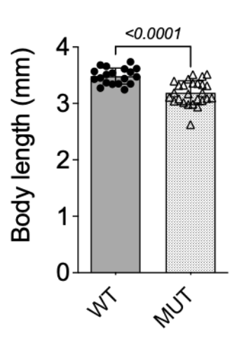
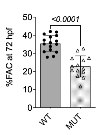
To gather information about the fish’s heart I organized data in Microsoft Excel. In an app called Fiji, I would measure the area of the heart in diastolic and systolic. From there I could measure the fraction area change (FAC) of the heart. In addition to measuring the heart, I also measured the fish’s length. I found that the mutant fish were smaller than the wild type. This meant that the foxc1a in the zebrafish aided with embryonic growth. I also found that mutant fish had a lower FAC which is a phenotype of HLHS. In conclusion, I found that a mutation in the FOXC2 gene can cause HLHS.
Graphs of data that I collected
In addition to working at the lab, I had the opportunity to attend Journal Club along with all the other interns in the cardiology department. Each week I would be assigned to read a scientific journal. I learned about many interesting topics such as heart regeneration, treatment for cardiac diseases, and heart development in babies. Journal Club was an amazing opportunity to practice my scientific text analysis skills along with learning about new and interesting topics.

I also had the opportunity to shadow Dr. Vassilio Bezzerides. I shadowed him through the intensive care unit and the cardiac care unit. There, I was able to watch him speak to patients and even watch an open-heart surgery. It was an eye-opening experience since I have been a patient many times at Boston Children’s but I have never been behind the scenes.
In my six weeks at the Burns Lab, I learned a great deal about the field of research. Specifically, I learned how to use lab equipment to run experiments and analyze data. In addition, I learned about the many different career paths in the world of STEM.

I would like to thank Dr. Caroline Burns, Dr. Geoff Burns, Dr. Mengmeng Huang, and the rest of the staff at the Burns Lab. From the beginning, everyone was so welcoming and patient. I appreciate the knowledge and answers to my constant questions. I had an amazing time and learned so much about research and cardiology.
Photo with the whole lab Me and both Doctor Burns


Comments